
Business Type:
Trading Company
Establishment:
2011
R&D Capacity:
OEM, ODM, Others
Terms of Payment:
LC, T/T, D/P, Paypal, Western Union
Main Markets:
North America, Europe
Guangzhou MeCan Medical Limited is a professional medical and laboratory equipment manufacturer and supplier. For more than ten years, we engage in supplying competitive price and quality products to ...
1
- YRS


 Dialysis Machine Crrt Dialysis Machine Crrt Hemo
 Video Gastroscope Gastroscope And Colonoscope En
 Portable Xray Machine Good Service For Mobile X-
 Portable Dialysis Machine Guangzhou Portable Dia
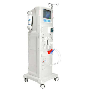 Equipment Peritoneal Dialysis Equipment Peritone